Datanami
 EnterpriseAI
 HPCwire Japan
 QCwire
 HPC & AI Wall Street
With the combined power of cryo-electron microscopy and high performance computing, scientists are opening up new frontiers in biochemistry.
In the fall of 2017, three distinguished scientists shared a Nobel Prize in Chemistry for pioneering a microscope technology that promises to revolutionize biochemistry. This technology, cryo-electron microscopy, or cryo-EM, opens the door to new levels of scientific discovery, including the visualization of proteins at a near-atomic level.
In simple terms, cryo-EM allows researchers to freeze biomolecules in mid-movement and produce three-dimensional structures of them. These 3D simulations help scientists visualize and understand how biomolecules function and interact — processes that would otherwise be impossible to see.
In announcing the award, the Royal Swedish Academy of Sciences noted that current scientific literature is filled with images of everything from proteins that cause antibiotic resistance to the surface of the Zika virus, and that “biochemistry is now facing an explosive development and is all set for an exciting future.”[1]
There are a wide range of use cases for cryo-EM. It is now seen as one of the keys to developing new pharmaceuticals and therapies, including innovations in cancer immunotherapy and precision medicine. The publication Chemistry World notes that, during an outbreak of the Zika virus in Brazil, a group of researchers used cryo-EM to generate a high-resolution 3D image of the virus structure, a view that provided a starting point in the search for sites that could be targeted by drugs to prevent the spread of the virus.[2]
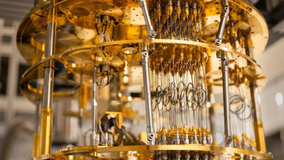
Cryo-EM alone, of course, doesn’t provide all the insights that researchers seek. Those insights come from the combination of cryo-EM and high performance computing (HPC) simulations, which churn through massive amounts of data to yield detailed 3D models of biological structures at sub-cellular and molecular scales.
Let’s look at a few examples of the way cryo-EM and HPC are serving as a catalyst for scientific discovery.
The Rockefeller University
At The Rockefeller University, the Evelyn Gruss Lipper Cryo-Electron Microscopy Resource Center makes sophisticated cryo-EM tools available to university researchers. These tools allow researchers to visualize the 3D structures of molecules and macromolecular complexes in solution.
Over the past four years, the use of cryo-EM has led to dozens of research breakthroughs at the university. In particular, cryo-EM has helped scientists understand the configurations and choreography of a range of previously intractable biological molecules. Some of these molecules are trademarks of deadly diseases, including cystic fibrosis, which make them important targets for new therapies.[3]
Peking University
At Peking University, researchers are leveraging the processing power of HPC clusters from Dell EMC to further cryo-EM cooperative research with Harvard University. These clusters, with 144 nodes and about 2 petabytes of storage with Lustre, enable researchers to map the 3D structure of biological macromolecules to design inhibitors and develop new drugs to treat or cure patients of cancer and other diseases.
“The HPC clusters from Dell EMC are critical to our research missions that highly depend on the analysis of big data generated from highly automated cryo-electron microscopes,” notes Dr. Youdong “Jack” Mao, assistant professor of biophysics at Peking University, in a news release highlighting the research effort. “The HPC systems facilitate the development of state-of-the-art algorithms in pursuit of structural solutions to those grand biomedical problems, which would deliver innovations in cancer immunotherapy and precision medicine.” [4]
Texas Advanced Computing Center
A team of researchers from four universities is using cryo-EM and supercomputing simulations run at Texas Advanced Computing Center (TACC) to model a vital molecular machine known as the human pre-initiation complex (PIC). The goal of this massive scientific investigation, which has involved millions of processor-core hours of simulations, is to produce atomic models that tell the full story of the structure and function of the protein complex of molecules.
A researcher on the project notes that this work lays the foundation for the development of future cures, which wouldn’t be possible without an understanding of the how PIC and other complex molecular machines function. [5]
Revolutionizing medical research
As examples like these show, the combination of cryo-EM and HPC is revolutionizing biochemistry and medical research. It is helping researchers make the fundamental scientific discoveries that create fertile ground for the development of life-saving pharmaceuticals and therapies, like immunotherapy and precision medicine.
And at the end of the day, that’s what really matters — saving lives.
For a closer look at the simulations made possible by cryo-electron microscopy and high performance computing, watch the Cryo-EM demo on Dell PowerEdge servers.
[1] Royal Swedish Academy of Sciences news release, “The Nobel Prize in Chemistry 2017,” October 4, 2017.
[2] Chemistry World, “Explainer: What is cryo-electron microscopy,” 2017.
[3] The Rockefeller University, “Third Rockefeller cryo-EM to help tame poorly behaved proteins,” August 30, 2018.
[4] Dell EMC news release, “Dell EMC Expands High Performance Computing Portfolio with Advances in Cloud, Software and System,” November 15, 2016.
[5] Texas Advanced Computing Center, “How To See Living Machines,” Published on November 21, 2016 by Jorge Salazar.