Datanami
 EnterpriseAI
 HPCwire Japan
 QCwire
 HPC & AI Wall Street
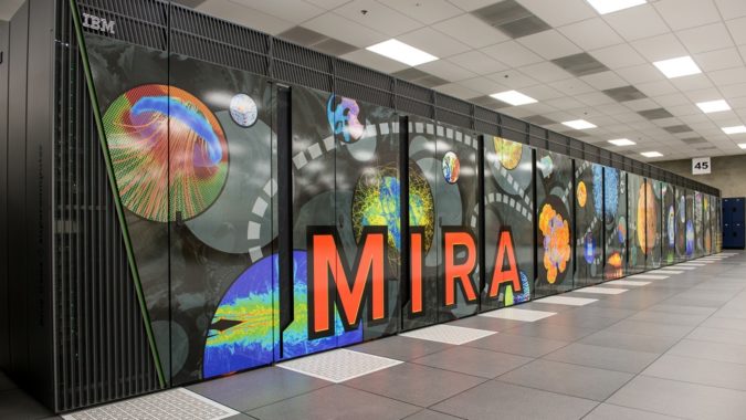
Reliable atomic-scale imaging of freeform molecules would unlock countless possibilities, particularly in fields like drug design and virology, where it could be used to quickly study the structure of viruses. Now, researchers at Argonne National Laboratory have applied supercomputer-powered simulations in an attempt to advance one method of atomic-scale molecular imaging.
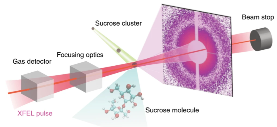
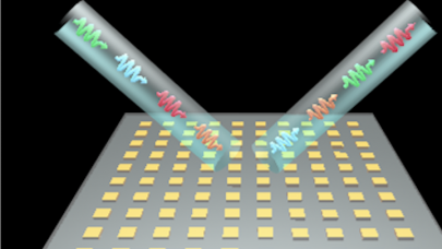
Currently, researchers derive atomic-scale models of molecules by crystallizing them and hitting them with x-rays, scattering the light and allowing for a pattern to be derived. Unfortunately, this method is less reliable with certain biological samples. One alternative method involves firing extremely fast, intense x-ray free-electron lasers (XFELs) at molecules, negating the need for crystallization. This technique works – but it has a cost.
“For this type of experiment, you need very intense pulses, which can destroy the sample very quickly,” said Phay Ho, an Argonne physicist and co-author of the research. “With this approach, you need to use very short pulses so you can collect all the scattering signals before the sample is destroyed.”
Working with XFELs for imaging of this kind requires temporal accuracy in the femtoseconds – 10-15 seconds – and spatial accuracy in nanometers. Beam time for real-world experiments, Ho explained, was difficult to obtain: so instead, the researchers decided to explore XFEL-molecule interactions through a series of molecular simulations covering 42 million particles.

To run these simulations, they turned to Argonne National Laboratory’s massive Mira supercomputer. Mira, an IBM Blue Gene/Q system, holds 49,152 nodes (each equipped with a 16-core IBM PowerPC A2 processor) and 768 TB of memory. In total, Mira delivers 8.59 Linpack petaflops, placing it 22nd on the most recent Top500 list of the world’s most powerful publicly ranked supercomputers.
“When you have a machine like Mira, you can run a large number of simulations, you can do them all at the same time, and you can run them over the timescales that we needed for this particular study,” said Christopher Knight, a computational scientist at Argonne and another co-author of the research.
The researchers found that time was even more critical than previously thought.
“If you use pulses this long, you can actually degrade your signal substantially,” Ho said. “In order to do this type of imaging, the pulse should last only a few femtoseconds. It’s important to look not just at the number of photons, but the number of photons per unit of time.”
With this new information in hand, the researchers are now better-prepared to approach the critical real-world experiments with their best foot forward.
To read Argonne’s Christina Nunez’s article discussing this research, click here.